A board-certified radiologist in Boston, Gul Moonis, MD, has extensive experience in the field of temporal bone imaging. Dr. Gul Moonis has published several articles on this topic, including a work on the anatomy of the temporal bone referencing MR and CT scanning methods published in the journal "Radiology".
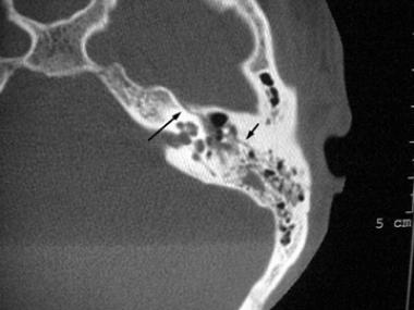
Recent advances in magnetic resonance (MR) technology have provided greater accuracy in diagnosing a diverse range of issues surrounding the temporal bone, from cholesteatoma to hearing loss. Temporal bone imaging through MR scanning focuses primarily on concerns related to soft tissue. For conditions affecting the temporal bone itself, computed tomography (CT) scanning provides greater structural detail. Both MR and CT scanning of the temporal bone assist in distinguishing different types of cancers, however.
Through either technology, temporal bone scanning offers an illuminating view into the five different parts of the temporal bone known as the styloid, tympanic, petrous, mastoid, and squamous portions, along with their integral pathways. Each of these areas tends toward distinct abnormalities which can be distinguished through the imaging process. Issues may result from chronic, or acute conditions.
 RSS Feed
