Gul Moonis, MD, serves as a neuroradiologist and diagnostic radiologist through Beth Israel Deaconess Medical Center. Acknowledged as one of America's Top Doctors by Castle Connolly, Dr. Gul Moonis draws on particular expertise in functional brain mapping.
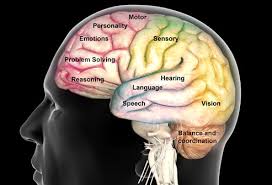
In every human brain, distinct areas control particular conscious and unconscious functions, including movement, language, and sensation. The exact location of these areas differ somewhat from person to person, which means that any kind of surgery involving access to the brain is extremely delicate. For this reason, patients who need surgery to remove damaged brain tissue often undergo functional brain mapping, which helps the surgical team to remove only the correct tissue.
Part of the mapping procedure takes place under general anesthesia. With the patient asleep, the medical team stimulates a nerve in one of the patient's limbs and records the neurological response. This is known as sensory mapping, or more specifically, somatosensory evoked potentials. The team also conducts motor mapping, a similar process in which electrical current stimulates the brain and causes movement of the body.
The team must then wake the patient to perform language mapping. This is done after the team has numbed the scalp, so that the patient will feel no pain.
Once awake, the patient must associate names with various images. The team stimulates his or her brain and finds the location at which the person can no longer name the object. This is an indication that the team has found the language center.
This process is often used to identify the site of excision for patients with brain tumors or seizure disorders. It may also serve as a diagnostic tool for certain neurodegenerative disorders, including Parkinson's and Alzheimer's diseases. In addition, some researchers use it to learn more about conditions such as autism, schizophrenia, and bipolar disorder
 RSS Feed
